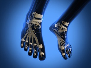
What Tests Are Used To Check for Osteoporosis?
Osteoporosis is usually symptom free until a fracture occurs, by which point it is more difficult to treat the osteoporosis or reduce the risk of bones thinning even further.
There are a number of tests available to diagnose osteoporosis, or help confirm diagnosis of other health conditions that may be causing bone loss.
DEXA Scan
Dual energy x-ray absorptiometry (DEXA or DXA) is the most common way to diagnose osteoporosis. The DEXA scan measures bone mineral density (BMD) in a number of areas of the skeleton, such as the hip, lower spine or wrist. The scan uses a low dose of radiation (around 10% of the dose from a chest x-ray). In a DEXA scan, or peripheral DEXA scan, you lie still on a couch for no more than twenty minutes, while scanning equipment moves over the body, taking an image. From this information, a T score is obtained. The T score is the comparison of your results against the range of young adults that are healthy and have an average bone density.
A T score of below -2.5 means that a diagnosis of osteoporosis is confirmed; a score of between -1 and -2.5 is diagnosed as osteopenia – some loss of bone density, but not enough to be diagnosed as osteoporotic. Individuals can often prevent osteopenia from developing into osteoporosis by making lifestyle changes such as staying active and eating a well-balanced diet. A T score of between 0 and -1 is considered normal bone density. A Z score is sometimes available; this compares your bone density to the density of people the same age as you.
DEXA scanning in the elderly can be difficult, as the spine may be affected by other disorders such as osteoarthritis. This means that some bones can appear denser during a scan than they actually are. Some DEXA scans may also incorporate vertebral fracture assessment or VFA, which allows spinal fractures to be detected more easily.
Physical Exam and Medical History
A physical examination for osteoporosis involves measuring height and checking the spine. Many professionals will also question you about risk factors such as your diet, how much you exercise, smoking and drinking habits, family history, fracture history and other factors that may contribute to your risk of developing osteoporosis. They will also take into account your DEXA scan results, or results from other bone tests.
FRAX® score
This is a fracture risk assessment tool developed by the World Health Organization. It includes bone density scores and other information to estimate your fracture risk for a ten year period. It is often used for those with osteopenia, those over the age of 50, or those who have yet to start treatment for osteoporosis.
Ultrasound
Quantitative ultrasound (QUS) can assess bone density in legs, heels and fingers. It uses sound waves to assess BMD, although unlike DEXA, it cannot assess bones such as the hip and spine – often the bones with the highest risk of osteoporotic fracture. A heel (calcaneus) ultrasound takes only a few minutes.
Other Tests
Depending on the results of some of the following tests, further investigations may be required to either confirm the diagnosis of osteoporosis, or confirm diagnosis of another condition that is causing bone loss or fractures.
The x-ray is the traditional diagnostic tool to check for fractures and may take place alongside a DEXA scan. X-rays are also useful for assessing height loss or curvature of the spine. Bone (radioisotope) scans use a dye which shows up on a scan, helping to identify fractures that an x-ray may have missed.
Additional tests such as blood calcium levels, urine calcium measurements and hormone levels can assist clinicians in identifying causes for bone loss, as there are some health conditions such as thyroid diseases that can cause secondary osteoporosis. Other lab tests, such as bone marker tests can also assess how fast bone is being lost or made.
A CT (computer tomography) scan may be available if there are problems measuring bone density, such as after a compression fracture in the spine. Although an MRI (magnetic resonance imaging) scan doe not measure bone density, it can be valuable in identifying spine or hip fractures.
Your clinician will usually discuss testing options with you, although for the majority of individuals, the DEXA scan is usually accurate enough to assess bone thinning and diagnose osteoporosis.
References
- Making a Diagnosis: <href=”#frax”>http://www.nof.org/articles/8#frax
- Scans and Tests and Osteoporosis: http://www.nos.org.uk/NetCommunity/Document.doc?id=388
Last Reviewed 11/Mar/2014
