
Breast MRI Scan Fact Sheet
Breast cancer is a leading cause of death among women. Regular screening tests can detect cancer in time to lower the risk of death or prevent the development of cancer when precancerous cells are present. Breast magnetic resonance imaging (MRI) scans are a type of test that uses radio waves and magnets to get images of the breast tissue without the need for x-rays. MRI scans are used in conjunction with mammograms and ultrasounds.
Overview of the Test
Breast MRI scans show clear images of areas of the breast that are not always accessible with mammograms and ultrasounds. These tests may find signs of cancer, reveal the existence of scar tissue and provide more information on a patient with an abnormal mammogram. MRI scans of the breast are also useful for analyzing a lump following biopsy, revealing breast implant ruptures and, in some cases, guiding a biopsy procedure. Scans show how well blood is circulating through the breasts and provide information about the blood vessels in the area.
Women who have thick breast tissue and are considered high risk for cancer of the breast are often given MRIs instead of mammograms.
Evidence and Science behind the Test
Multiple studies show that MRIs are useful as a screening tool for breast cancer, particularly in women who are high risk candidates. A 2004 study published in the New England Journal of Medicine compared the ability of mammograms to detect cancer with that of MRIs. The mammograms found 18 cancers and missed 22 that MRIs caught. The MRIs detected 32 instances of cancer and missed eight that mammograms found.
A 2007 study published in Radiology journal showed comparable results. The participants were given MRIs as well as mammograms, ultrasounds and clinical examinations. The MRI detected 17 of 18 cancers, and six of those had been missed by all the other screenings.
Another study the same year showed that MRI scans found all six cancers developing in the study’s participants. Mammograms caught only two of the six and an ultrasound discovered one. The study indicated that only MRIs were capable of finding cancer in dense tissue.
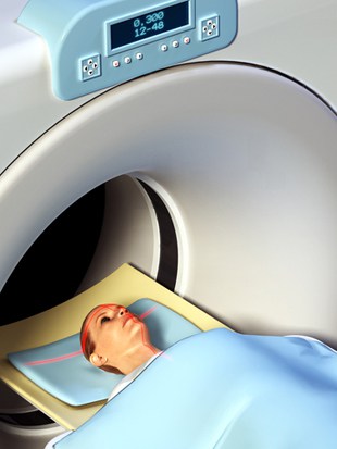
How is it done?
A MRI scan is a relatively simple procedure. Patients must take off any metal, such as earrings beforehand. A healthcare professional insets a catheter into the patient’s arm. The staff member injects gadolinium into the arm prior to the MRI, or sometimes during.
The patient lies face down on a table with their breasts in a cushioned recess. Inside the recess is a signal receiver. The table slides into the scanner. The patient stays still for 15-minute intervals while the machine takes images. The complete procedure usually takes 30 to 60 minutes. Sometimes, these scans take longer.
Who Does It?
Doctors.
When and How Often?
Women whose risk of breast cancer is more than 20 percent should have their breasts scanned every year. Women with a 15 percent risk or higher may also need yearly scanning, but should discuss this with their doctors first.
Cost
A breast MRI scan costs more than $1,000 and may be more than $2,000. Most insurance companies do not cover the cost of a breast MRI. Some companies do cover high-risk individuals, and these people may pay little or no money out of pocket for the test.
Issues
Although MRI breast scans offer clearer pictures than mammograms and can detect cancer in dense tissue that other methods might not catch, the images are almost too good. They may detect abnormalities that are not cancerous.
References
Last reviewed 26/Feb/2014


